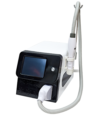
Picosecond Laser Technology: Clinical Evidence and Emerging Applications
2026-09-09
Introduction
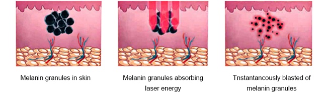
Picosecond lasers deliver energy in pulses measured in trillionths of a second—300 to 750 picoseconds in most clinical systems. Compared to conventional nanosecond Q‑switched lasers, picosecond pulses generate substantially higher instantaneous peak power, producing a dominant photoacoustic (photomechanical) effect on target chromophores with significantly less thermal diffusion to surrounding tissues. This fundamental difference in pulse duration translates to more efficient pigment fragmentation, reduced collateral thermal damage, and a lower risk of post‑inflammatory dyspigmentation—particularly relevant for patients with skin of color.
The clinical literature on picosecond laser technology has expanded considerably in recent years, with multiple randomized controlled trials and systematic reviews published between 2025 and 2026. This article synthesizes the current evidence on picosecond laser applications across pigmentary disorders, acne scarring, skin rejuvenation, and emerging indications, with emphasis on data from well‑designed comparative studies.
Atrophic Acne Scarring: Comparative Effectiveness
Acne scarring remains one of the most common indications for fractional laser therapy. A prospective, randomized split‑face study published in 2026 compared the efficacy and safety of 1064‑nm picosecond laser with 1927‑nm fractional thulium laser for atrophic acne scars in Asian patients. Both modalities demonstrated comparable clinical efficacy, suggesting that picosecond laser is a viable alternative to established fractional thulium systems for scar revision in Asian skin types.
A separate 20‑week prospective, randomized split‑face study compared 755‑nm picosecond laser with diffractive lens array (P‑DLA) against 1565‑nm nonablative fractional laser (NAFL) for atrophic acne scars. Both P‑DLA and NAFL were effective and safe modalities. However, a key mechanistic distinction emerged: NAFL primarily targets water through fractional photothermolysis, creating microthermal zones in the dermis that trigger collagen remodeling, whereas P‑DLA interacts primarily with epidermal melanin and may induce laser‑induced optical breakdown (LIOB) within the epidermis. The resulting pressure waves from P‑DLA propagate into the dermis and may influence cellular signaling and promote dermal remodeling. Histological studies of picosecond lasers have reported that the stratum corneum and surrounding tissues remain intact with no evidence of thermal injury. This difference partially accounts for the reduced downtime and absence of crusting observed with P‑DLA.
For patients prioritizing minimal downtime and fewer adverse effects, P‑DLA may be more suitable. The absence of crusting and reduced recovery time represent meaningful clinical advantages, particularly for patients with limited tolerance for post‑procedure social downtime.
Pigmentary Disorders: New Data on Wavelength Selection Melasma
Melasma remains one of the most challenging pigmentary disorders to treat, with high recurrence rates and significant risk of post‑inflammatory hyperpigmentation (PIH), particularly in darker skin types. The pathological changes in melasma extend beyond the epidermis to include dermal findings such as melanophages, perivascular lymphocytic infiltration, solar elastosis, and vascular proliferation.
A 2025 systematic review and meta‑analysis of randomized controlled trials evaluating 755‑nm picosecond alexandrite laser (PSAL) for melasma found low rates of irreversible adverse events. However, the meta‑analysis also reported that PSAL appeared inferior to triple combination cream in reducing Melasma Area and Severity Index (MASI) scores, and post‑inflammatory hyperpigmentation was more common in the PSAL group compared to topical creams (OR = 6.86, 95% CI: 1.47‑32.07). There was no significant difference in PIH rates when PSAL was compared with Q‑switched lasers. No incidence of hypopigmentation or infection was reported.
A randomized evaluator‑blinded trial published in 2025 directly compared a novel picosecond alexandrite laser (PSAL) with traditional combined Q‑switched and long‑pulse Nd:YAG lasers (QLNYL) in 40 participants with Fitzpatrick skin types III‑IV. Both groups experienced significant reductions in MASI scores post‑treatment. However, the improvement in MASI scores in the QLNYL group significantly surpassed that in the PSAL group (P = 0.010). Patient satisfaction was comparably high between groups, and no significant differences were noted in safety profiles. The PSAL group showed a slightly higher incidence of adverse reactions (not significant) and significantly higher pain scores (P = 0.018). Recurrence rates at 24‑week follow‑up were 10.5% for PSAL and 0% for QLNYL, with no significant difference.
A prospective single‑group study evaluated a 785‑nm picosecond titanium:sapphire laser for melasma in Asian patients. After 10 treatment cycles with 2‑week intervals, independent dermatologists scored significant improvement at week 10 (0.97 ± 0.51, slight improvement), with further improvement at week 22 (1.44 ± 0.81, moderate improvement). The percent improvement was 12.33% ± 9.78% at week 10 and 22.97% ± 15.40% at week 22, and general skin color and diffuse erythema also improved.
Low‑fluence 730‑nm picosecond laser has also emerged as an effective and safe modality for melasma in Chinese patients. The 730‑nm wavelength offers advantages in melanin selectivity and relatively deep penetration while maintaining a short pulse width. Acquired Dermal Melanocytosis (Hori's Nevus)
A retrospective study comparing 730‑nm and 1064‑nm picosecond lasers for acquired dermal melanocytosis (ADM) found that after three sessions, the 730‑nm group exhibited superior outcomes in terms of subjective symptoms (patient satisfaction), objective findings, and software analysis improvements. The incidence of hyperpigmentation was 15.4% in the 1064‑nm group and 14.5% in the 730‑nm group, with no significant differences in complication rates. The authors concluded that the 730‑nm picosecond laser, which possesses high melanin selectivity, relatively deep penetration, and a short pulse width, suggests a potentially more effective treatment for ADM compared to the conventional 1064‑nm wavelength, without increasing complications. Lichen Planus Pigmentosus
Fractional picosecond Nd:YAG laser has also been evaluated for lichen planus pigmentosus (LPP), a chronic dermal pigmentary disorder predominantly affecting darker skin types. In a prospective randomized study of 20 Indian patients, Group A received four sessions of fractional 1064‑nm picosecond Nd:YAG laser followed by toning every 4‑6 weeks, while Group B underwent laser toning alone. Group A showed superior pigment clearance with a mean reduction of 36.2% vs. 21.8% in Group B. Sixty percent of Group A achieved >75% clinical improvement compared to only 10% in Group B. Patient satisfaction scores were higher in Group A (6.7 vs. 4.6). No long‑term adverse effects were observed.
Skin Rejuvenation and Photoaging
A prospective randomized trial comparing 1064‑nm fractional picosecond laser with intense pulsed light (IPL) for facial rejuvenation in Asian women found both modalities effective and safe. The picosecond laser's photoacoustic mechanism, minimal thermal damage, and potential for deeper dermal remodeling make it a promising tool for skin rejuvenation.
Histological evidence confirms that picosecond laser treatment stimulates collagen formation. Collagen formation is most effective using 1064‑nm pulse duration of 400 ps, with the effect increasing proportionally with laser power. In vitro studies have demonstrated that low‑energy picosecond laser pulses can penetrate deeply into the dermis, inducing laser‑induced cavitation and promoting collagen regeneration. Confocal microscopy has confirmed collagen remodeling and formation of new dermal papillae following fractional picosecond laser treatment.
A split‑face study comparing fractional 1064‑nm Nd:YAG picosecond laser with fractional Q‑switched 1064‑nm Nd:YAG laser in photoaging improvement found that the picosecond laser modulates the expression levels of collagen, TIMP, and MMP by downregulating miR‑24‑3p, thereby fostering collagen synthesis and fortifying protective effects on the skin barrier.
Safety in Skin of Color: A Systematic Review
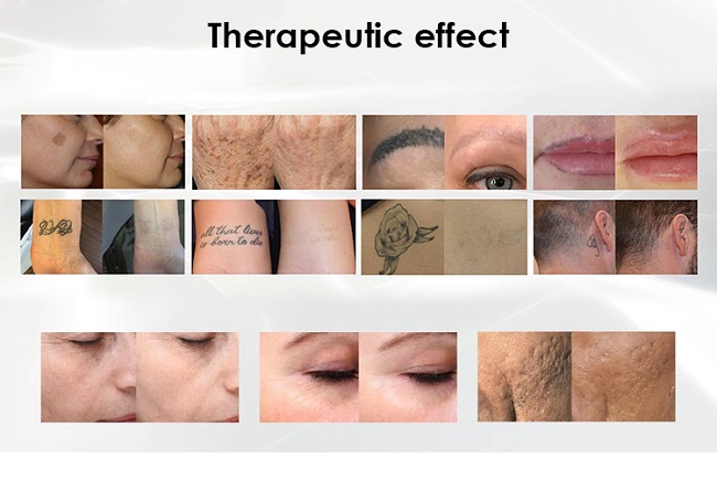
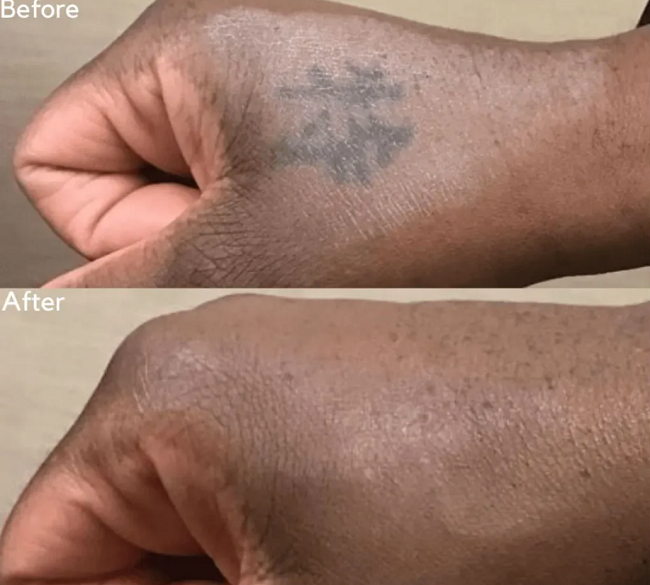
A 2026 systematic review evaluated the safety and efficacy of picosecond laser therapy in patients with Fitzpatrick skin types IV‑VI. Among 257 patients across 21 studies, PSL demonstrated moderate to excellent improvement across pigmentary and nonpigmentary conditions, including acne, scarring, photoaging, striae distensae, and lichen amyloidosis. Adverse effects were generally mild and transient, with low rates of dyspigmentation and no permanent scarring. Few comparative studies suggested superior pigment clearance and reduced PIH with PSL compared with nanosecond lasers.
A meta‑analysis comparing picosecond versus nanosecond lasers for hyperpigmented disorders and tattoos found that adverse events including PIH and post‑inflammatory hypopigmentation were much more obvious in Asian than European patients. Compared with nanosecond lasers, picosecond lasers had significantly lower rates of PIH (P = 0.02). Another split‑face study comparing 532‑nm picosecond and Q‑switched Nd:YAG lasers for solar lentigines reported a PIH incidence of 5% in the picosecond laser‑treated sides and 30% in the Q‑switched laser‑treated sides.
Combination Therapy Approaches
Combination strategies are increasingly being explored to enhance picosecond laser outcomes. Low‑fluence 1064‑nm picosecond laser with micro‑lens arrays (MLA) improved melasma severity and reduced recurrence rates compared with low‑fluence picosecond laser alone.
In tattoo removal, the addition of radial shock acoustic wave therapy to picosecond laser treatment appears to offer a more effective and well‑tolerated approach. The R20 method (repeated passes with short intervals) with picosecond laser has also shown the most significant improvement in clinical evaluation.
Fractional and non‑fractional picosecond Nd:YAG lasers combined with fractional picosecond KTP laser for melasma in Chinese patients showed significantly better efficacy than using single wavelength fractional and non‑fractional picosecond Nd:YAG lasers alone.
Emerging Applications
A 532‑nm picosecond laser has been evaluated for depigmentation therapy of refractory facial vitiligo. In two case reports, the 532‑nm picosecond laser, followed by topical hydroquinone, appeared to be an effective and well‑tolerated approach for managing residual pigmentation in vitiligo patients. Marked reduction in residual pigmentation was observed in both patients, with cosmetically favorable outcomes.
Fractional 1064‑nm picosecond laser has also been studied for post‑mammoplasty scar minimization in Asians and for abdominal striae alba, demonstrating safety and efficacy in these emerging applications. Post‑acne erythema has also been effectively treated with 1064‑nm picosecond laser with micro‑lens array.
Conclusion
The clinical evidence base for picosecond laser technology has expanded substantially, with multiple randomized controlled trials and systematic reviews published in 2025‑2026. The technology demonstrates robust efficacy across pigmentary disorders, acne scarring, and skin rejuvenation, with a favorable safety profile that extends to Fitzpatrick skin types IV‑VI.
Key takeaways from recent evidence:
Picosecond lasers achieve comparable efficacy to fractional thulium systems for atrophic acne scars with reduced downtime
Wavelength selection is critical: 730‑nm shows superior outcomes for acquired dermal melanocytosis; 785‑nm demonstrates efficacy in Asian melasma
Combination approaches—particularly with low‑fluence protocols and adjunctive therapies—enhance outcomes
The safety profile in skin of color is favorable, with low rates of dyspigmentation and no permanent scarring reported in systematic reviews
As with any energy‑based procedure, outcomes depend on appropriate patient selection, correct wavelength and parameter selection, and practitioner expertise. The expanding evidence base supports picosecond laser technology as a versatile and increasingly well‑validated tool in aesthetic dermatology.